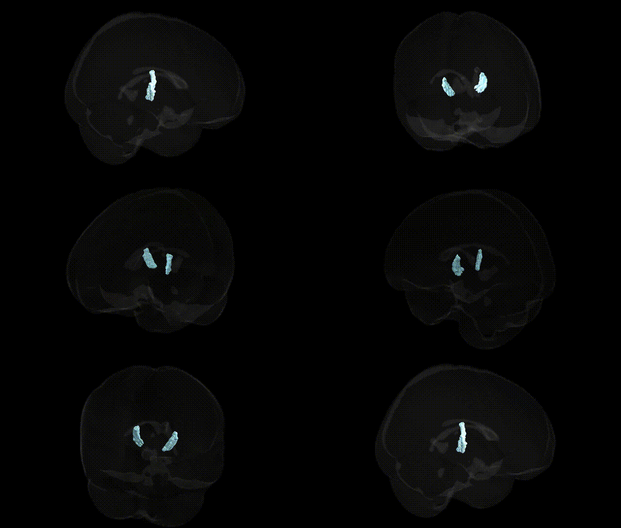
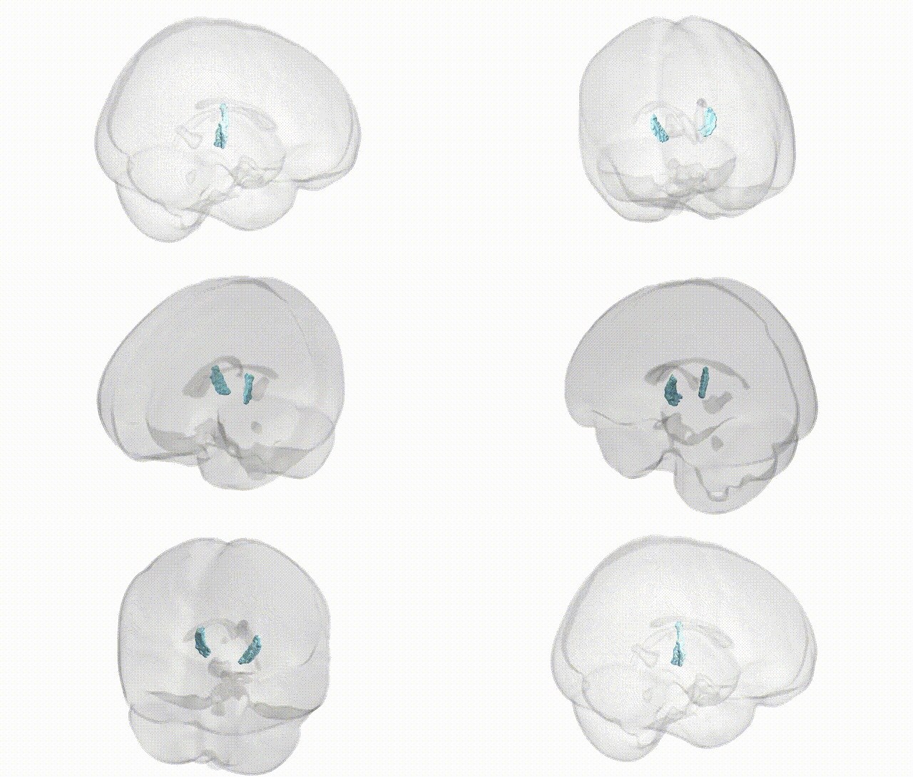
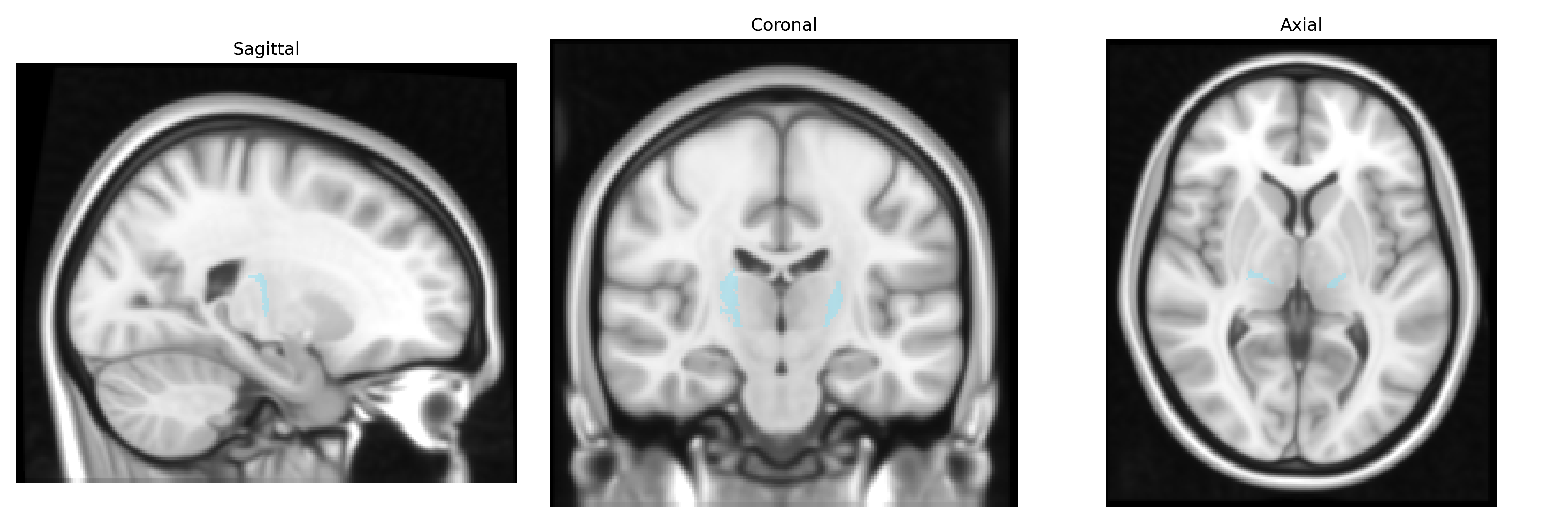
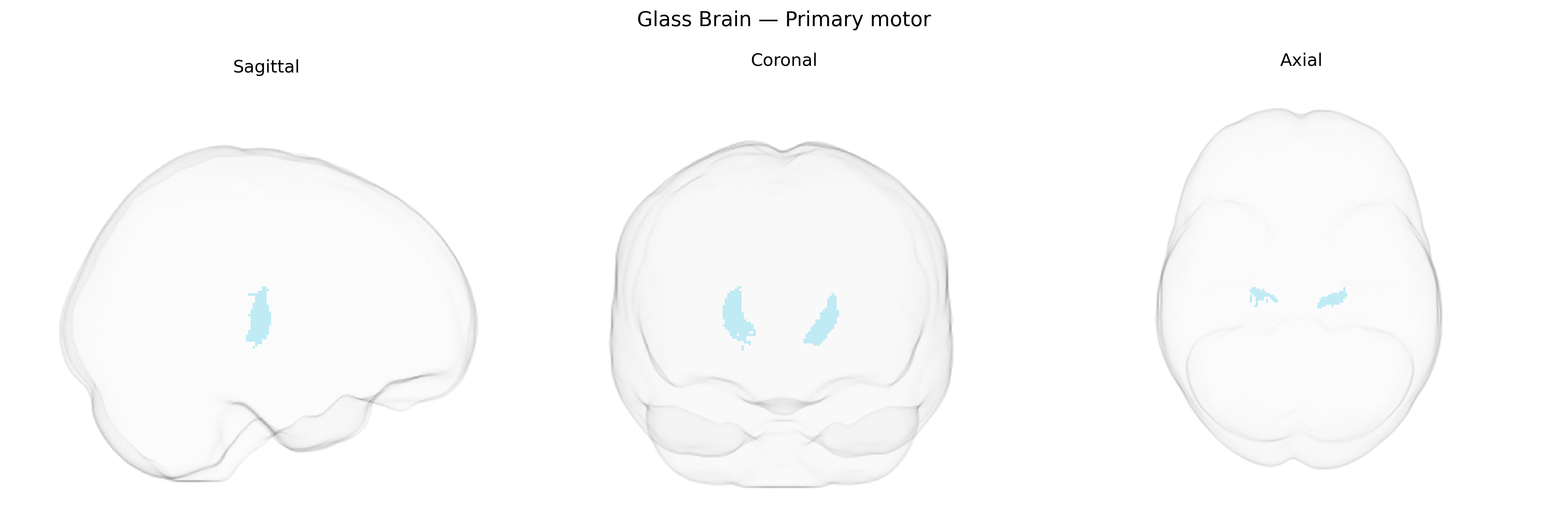
The bilateral Primary motor thalamic region (Thalamus maxprob thr25 1mm Atlas) corresponds predominantly to ventrolateral and ventroanterior nuclei that relay cerebellar and basal ganglia output to the primary motor cortex, forming a key node in cortico-subcortical motor circuits. Neurons in this region integrate excitatory and inhibitory inputs from the cerebellum, globus pallidus, and other motor-related structures, then project topographically to motor cortical areas to modulate voluntary movement initiation, execution, and muscle tone. Functionally, this thalamic motor territory participates in refining motor commands, coordinating agonist–antagonist muscle activity, and contributing to motor learning and adaptation. Lesions or dysfunction in this region are associated with movement disorders such as tremor, dystonia, and parkinsonian motor symptoms, reflecting its critical role in normal motor control. There is no direct Wikipedia article for this specific atlas-defined region; a closely related structure is the Ventral anterior nucleus of thalamus.
The bilateral primary motor thalamic region (often encompassing ventrolateral and ventroanterior nuclei projecting to primary motor cortex) has been implicated in genetic studies predominantly through GWAS and imaging–genetics work linking thalamic structure and thalamo-cortical circuits to neurodevelopmental and neuropsychiatric traits. Variants in genes affecting synaptic plasticity, axon guidance, and neurodevelopment (for example, CACNA1C, GRIN2B, CNTNAP2, and BDNF) have been associated with altered thalamic volume or connectivity to motor areas, and with disorders featuring motor or psychomotor abnormalities, including schizophrenia, autism spectrum disorder, ADHD, and major depressive disorder. Polygenic risk scores for Parkinson’s disease and essential tremor show associations with altered activity and connectivity in motor thalamic pathways, consistent with the role of this region in motor control; similarly, risk variants in genes related to dopaminergic and basal ganglia–thalamocortical function (such as LRRK2 and SNCA for Parkinson’s disease) map onto circuits that include the motor thalamus. Imaging–genetics studies also link common variants influencing intracranial and subcortical volumes (for example in HMGA2 and other growth-related loci) to individual differences in thalamic morphology, and GWAS of motor-related traits—such as reaction time, gait parameters, or motor learning—often highlight loci involved in cortical–subcortical connectivity, indirectly implicating the primary motor thalamus as a relay node within genetically influenced motor networks rather than identifying region-specific “motor thalamus genes.”
Overview generated by GPT-4o (2026).
Region ID: 1
Hemisphere: bilateral
Atlas: Thalamus maxprob thr25 1mm
Full Quality Version: Download MP4
Full Quality Version: Download MP4
Wali Sidiqyar*, Gaurav Rudravaram*, Elyssa M. McMaster, Trent M. Schwartz, Adam M. Saunders, Kurt G. Schilling, Bennett A. Landman "Introducing SPINS: A Shared Public Visualization Library of Neuroanatomical Structures." Medical Imaging with Deep Learning- short paper
This resource is licensed under CC0 1.0 Universal (Public Domain).